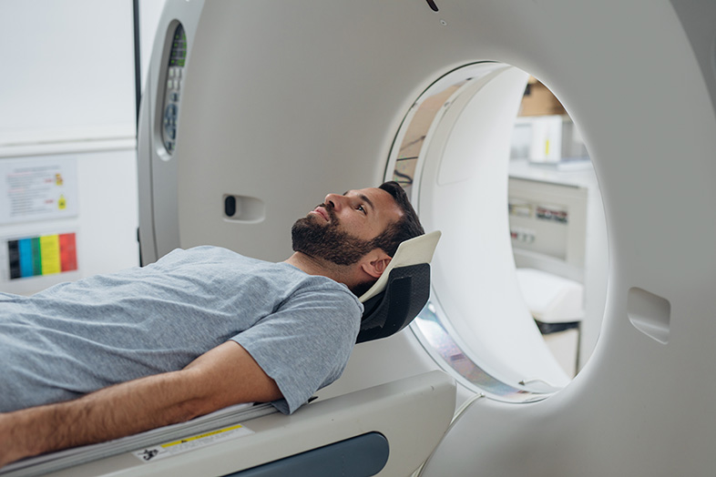
Suffering injuries in a car accident can be a traumatic event, but when those injuries cannot be seen by the naked eye, such as those suffered to the brain and head, it can make things that much worse. While CT and normal MRI scans can offer doctors a look into the human body, they often times do not show the extent of damage received, which can lead to questions of long-term effects.
But researchers believe that they may have found a better way to diagnose mild traumatic brain injuries in patients even years after an incident has occurred. With their new imaging technique, scientists are able to not only show where a person received damage in their brain but can make a prediction about how it will affect the patient down the road.
Called diffusion tensor imaging, also called DTI, the new imaging method identifies injuries in a person's brain by showing how fluid moves across the nerve fibers in the brain. Using a measurement called fractional anisotropy, scientists were able to measure how quickly fluid travelled over healthy nerve fibers versus damaged ones. Using veterans who had been diagnosed with mild traumatic brain injuries and healthy soldiers, scientists were not only able to detect damage in the brain but were able to identify patients who had greater brain damage from those who had little damage.
Although the study has mainly focused on soldiers who have suffered brain damage due to a blast injury, many of our Wisconsin readers may realize the impact this new scanning technique could have on car accident victims as well. Identifying severe damage within hours of receiving an injury could mean access to better treatments and can help physicians provide better long-term health care as well in the future.
Source: MedicalNewsToday.com, "MRI shows long-term effect of blast-induced brain injuries," Catharine Paddock, PhD, Dec. 2, 2013
